© Universität Bielefeld


Forschung
- Changes in sphingolipids in toxic metal-induced oncogenic transformation
Project member(s): Wing-Kee
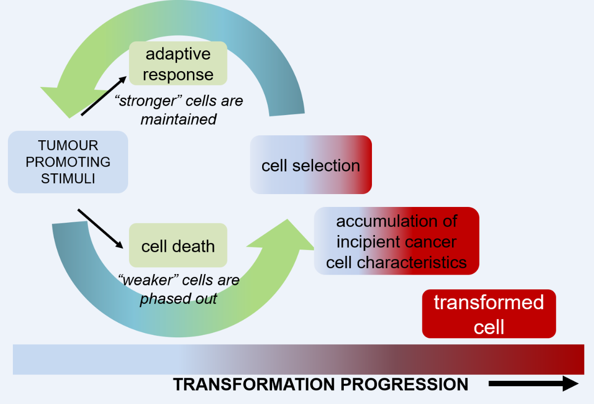
Heavy metals are omnipresent at low levels due to their presence in the earth’s crust but are released at potentially harmful levels into the environment. Cadmium and nickel and their compounds have been identified at class I carcinogens by the International Agency for Research on Cancer (IARC) and particularly target the kidney, wherein they accumulate. In the long-term process of oncogenic transformation of normal cells, it is hypothesized that carcinogens initiate cell selection whereby weaker cells are removed by cell death and stronger cells engage an adaptive response permitting cell survival despite harboring oncogenic mutations. As the adapted cells populate, oncogenic mutations accumulate and eventually give rise to cells with tumorigenic properties.

Abbildung: Wing-Kee Lee Previous work has evidenced cadmium and nickel-induced upregulation of the multidrug resistance P-glycoprotein ABCB1, a physiological efflux transporter that eliminates xenobiotics and toxic metabolic products and is expressed in the kidney, in the adaptive response. Importantly, changes in sphingolipid balance, in particular sphingomyelin and ceramide, appear to be an early event in carcinogenesis.
Current objectives aim to delineate sphingolipid changes at initial and late stages of carcinogenesis in the kidney by cadmium and nickel salts using in vitro and in vivo models and state-of-the-art cellular and molecular biology techniques to assess sphingomyelin/ceramide balance, alteration of sphingolipid enzyme activities, upstream signals (e.g. reactive oxygen species, calcium), and changes in lipid composition will be explored.
Collaborators:
Prof. Elmar Prenner (University of Calgary, Canada)
Prof. Frank Thévenod (Witten/Herdecke University)
Prof. Adriana Buzatto (University of Calgary, Canada)
Relevant selected publications
Lee, W.K., Probst, S., Santoyo-Sanchez, M.P., Al-Hamdani, W., Diebels, I, von Sivers, J.K., Kerek, E., Prenner, E.J., Thévenod, F. (2017) Initial autophagic protection switches to disruption of autophagic flux by lysosomal instability during cadmium stress accrual in renal NRK-52E cells. Arch. Toxicol. 91: 3225-3245.
Nair, A.R., Lee, W.K., Smeets, K., Swennen, Q., Sanchez, A., Thévenod, F., Cuypers, A. (2015) Glutathione and mitochondria determine acute defense responses and adaptive processes in cadmium-induced oxidative stress and toxicity of the kidney. Arch. Toxicol. 89: 2273-89
Dahdouh, F., Raane, M., Thévenod, F., Lee, W.K. (2014) Nickel-induced cell death and survival pathways in cultured renal proximal tubule cells: Roles of reactive oxygen species, ceramide and ABCB1. Arch. Toxicol. 88: 881-92.
Lee, W.K., Torchalski, B., Kohistani, N., Thévenod, F. (2011) ABCB1 protects kidney proximal tubule cells against cadmium-induced apoptosis: Roles of cadmium transport and ceramide metabolism. Toxicol. Sci. 121: 343-56.
Lee, W.K., Torchalski, B., Thévenod, F. (2007) Cadmium-induced ceramide formation triggers calpain-dependent apoptosis in cultured kidney proximal tubule cells. Am. J. Physiol. Cell Physiol. 293: C839-47.
Lee, W.K., Abouhamed, M., Thévenod, F. (2006) Caspase-dependent and –independent pathways for cadmium-induced apoptosis in cultured kidney proximal tubule cells. Am. J. Physiol. Renal Physiol. 291: F823-32.
- Cell organelles as targets of cadmium
Project members: Nadiya, Kaya, Bruce, Millie, Marie, Wing-Kee
Lipid membranes are used by cell organelles to form complex tightly regulated compartmentalized networks with specialized functions, which are fundamental to life. Interorganellar communication is crucial to orchestrate correct cell behavior, such as adaptive stress responses, and can be mediated by release of signaling molecules, exchange of organelle contents, mechanical force through organelle shape changes or direct membrane contact sites.
Cadmium, which once released into the environment cannot be further degraded, has become a major concern for public health and has been listed as one of the top 20 hazardous substances. Based on its chemical properties, cadmium uses molecular mimicry to move between cellular compartments, interacts with negatively-charged molecules (e.g. phospholipids), competes or displaces at metal-binding sites (e.g. on enzymes), and thereby affecting a plethora of cellular processes. As a result, organellar function is strongly impacted. Our previous work has focussed on mitochondrial uptake of cadmium and perturbance of mitochondrial function, autophagosome disruption and lysosomal instability by cadmium. Current projects examine the cadmium-cardiolipin connection, mitochondrial dynamics controlled by septins under cadmium, lysosomal cadmium-calcium interactions and cadmium impact on peroxisomes.
Collaborators:
Prof. Elmar J. Prenner (University of Calgary, Canada)
Prof. Frank Thévenod (Witten/Herdecke University, Germany)
Prof. Sven Thoms (Biochemistry and Molecular Medicine, Bielefeld University, Germany)
Relevant publications
Thévenod, F., Lee, W.K., Garrick, M.D. Iron and cadmium entry into renal mitochondria: Physiological and toxicological implications. Front. Cell. Dev. Biol. 8: 848. doi 10.3389/fcell.2020.00848.
Lee, W.K., Thévenod, F. (2020) Cell organelles as targets of mammalian cadmium toxicity. Arch. Toxicol. 94: 1017-1049. doi: 10.1007/s00204-020-02692-8
Lee, W.K., Probst, S., Santoyo-Sanchez, M.P., Al-Hamdani, W., Diebels, I, von Sivers, J.K., Kerek, E., Prenner, E.J., Thévenod, F. (2017) Initial autophagic protection switches to disruption of autophagic flux by lysosomal instability during cadmium stress accrual in renal NRK-52E cells. Arch. Toxicol. 91: 3225-3245. doi: 10.1007/s00204-017-1942-9
Lee, W.K., Bork, U., Gholamrezaei, F., Thévenod, F. (2005) Cd2+-induced cytochrome c release in apoptotic proximal tubule cells: role of mitochondrial permeability transition pore and Ca2+ uniporter. Am. J. Physiol. Renal Physiol. 288: F27-39. doi: 10.1152/ajprenal.00224.2004
- Regulation of ABCB1 by sphingolipids in cancer multidrug resistance
Project member(s):
Effective cancer chemotherapy treatment is often hampered by the development of multidrug resistance (MDR), which is in part defined by upregulation of ABCB1, a drug transporter, which extrudes chemotherapeutic drugs. Sphingolipids, such as ceramide and sphingomyelin, play multiple roles in the conferment of MDR by governing ABCB1 expression and activity. The lipid microenvironment affects ABCB1 functionalization; depletion of cholesterol and sphingomyelin, present in defined lipid domains called lipid rafts, leads to diminished ABCB1 transport activity. Furthermore, the balance between long-chain ceramides and very long-chain ceramides is altered in the MDR phenotype, possibly to support the functionalization of large drug transporters in the membrane, wherein ceramide synthases are involved.
In addition to drug extrusion at the plasma membrane, ABCB1 is mislocalized to an intracellular acidic vesicular pool wherein it sequesters chemotherapeutic drugs, further preventing attainment to their nuclear target. The sphingolipid ceramide increases vesicular fusogenicity and can initiate trafficking of ABCB1-drug containing vesicles to the nucleus, where the drug is deposited and kills cancer cells.
Current and future directions aim to isolate and characterize the molecular nature of the ABCB1-containing vesicular pool as well as decipher the role and impact of very long-chain ceramides and sphingomyelins in MDR in both tumor cells and cancer stem cells, which are particularly chemoresistant.
Collaborators:
Prof. Anthony H. Futerman (Weizmann Institute of Science, Israel)
Prof. Richard Kolesnick (MSKCC, USA)
Prof. Timan Kottke (Biophysical Chemistry and Diagnostics, Bielefeld University, Germany)
Relevant selected publications
Lee, W.K., Kolesnick, R.N. (2017) Sphingolipid Abnormalities in Cancer Multidrug Resistance: Chicken or Egg? Cell. Signal. 38: 134-145.
Dahdouh, F., Raane, M., Thévenod, F., Lee, W.K. (2014) Nickel-induced cell death and survival pathways in cultured renal proximal tubule cells: Roles of reactive oxygen species, ceramide and ABCB1. Arch. Toxicol. 88: 881-92.
Lee, W.K., Chakraborty, P.K., Thévenod, F. (2013) Pituitary homeobox 2 (PITX2) protects renal cancer cell lines against doxorubicin toxicity by transcriptional activation of the multidrug transporter ABCB1. Int. J. Cancer 133: 556-67.
Lee, W.K., Torchalski, B., Kohistani, N., Thévenod, F. (2011) ABCB1 protects kidney proximal tubule cells against cadmium-induced apoptosis: Roles of cadmium transport and ceramide metabolism. Toxicol. Sci. 121: 343-56.
- Lipocalin-2 and its receptor in cancer intercellular communication
Project member(s): Wing-Kee
Renal cell carcinomas (RCCs) are usually detected at advanced stages resulting in <10% survival rates and warranting development of new diagnostic methods. The iron-sequestering protein lipocalin-2 (LCN2) and its cell-surface receptor (LCN2R) are overexpressed in cancer, including colon, bladder and kidney, presumably to supply cancer cells with iron for metabolic requirements, growth and proliferation.
Cancer cells utilize extracellular messengers, such as RNA or protein, not only for tumor microenvironment maintenance but also to “educate” surrounding non-cancerous cells. Overexpression of LCN2 has been observed in tumor tissues, is postulated to contribute to cancer progression, drug resistance and metastatic potential and has been linked with poor prognosis. Secreted LCN2 from tumor cells appears to promote tumor progression through a number of mechanisms, including stabilization of matrix metalloprotease 9 permitting remodeling of the extracellular matrix and metastasis of tumor cells. In addition LCN2 captures iron, which is required for cell proliferation and tumor growth.
The interrelationship between secreted LCN2 and LCN2R in oncogenic transformation is a current research focus. Renal cancer cell lines show increased secretion of both LCN2 and LCN2R, which are released in an exosome-independent manner, compared to non-cancer renal cells. We are exploring the mechanisms by which LCN2 and LCN2R secretion impact transformation, inflammation and carcinogenesis of non-transformed renal cells using both 2D cell lines and 3D kidney organoids.
Collaborators:
Dr. M. Laura Martin (Altos Labs, San Francisco, CA)
Prof. Frank Thévenod (Witten/Herdecke University)
Relevant selected publications
Probst, P., Scharner, B., McErlean, R., Lee, W.K., Thévenod, F. (2019) Inverse Regulation of Lipocalin-2/24p3 Receptor/SLC22A17 and Lipocalin-2 Expression by Tonicity, NFAT5/TonEBP and Arginine Vasopressin in Mouse Cortical Collecting Duct Cells mCCD(cl.1): Implications for Osmotolerance. Int. J. Mol. Sci. 20: pii: E5398.
Langelueddecke, C., Roussa, E., Fenton, R.A., Wolff, N.A., Lee, W.K., Thévenod, F. (2012) Lipocalin-2 (24p3/Neutrophil Gelatinase-associated Lipocalin (NGAL)) receptor is expressed in distal nephron and mediates protein endocytosis. J. Biol. Chem. 287: 159-169.
- Ferroptosis resistance in tumor progression
Project members: Marie, Daniel, Teresa, Wing-Kee
Life is a delicate balance between order and disorder; one small disturbance is all it takes. The core concept of physiology – homeostasis – particularly resonates in the balance of cell death, a physiological process, which can be detrimental when unregulated. Ferroptotic cell death was discovered in 2012 by Brent Stockwell’s group and participates in tumor suppression, development and aging yet is strongly implicated in a number of diseases, including neurodegeneration and cancer.
Ferroptosis is a mode of programmed cell death dependent on iron (Fe) that is characterised by a defective antioxidant system, elevated membrane polyunsaturated fatty acids and lipid peroxidation. It is elicited by Fe-induced generation of reactive oxygen species (ROS), resulting in membrane damage and cell death. Though cancer cells are particularly susceptible to ferroptosis, recent data (Greene, C.J. et al. Front. Oncol. 12:923043, 2022) suggest renal cancers accumulate less iron with pathological progression, thus decreasing ferroptosis susceptibility. We are currently using different cancer cell line models to delineate the mechanisms of ferroptosis resistance.
Relevant publications
Thévenod, F., Lee, W.K., Garrick, M.D. Iron and cadmium entry into renal mitochondria: Physiological and toxicological implications. Front. Cell. Dev. Biol. 8: 848. doi 10.3389/fcell.2020.00848.
Smith, C.P., Lee, W.K., Hadley, M., Poulsen, S.B., Thévenod, F., Fenton, R.A. (2019) Proximal tubule transferrin uptake is modulated by cellular iron and mediated by apical membrane megalin-cubilin complex and transferrin receptor 1. J. Biol. Chem., 294:7025-7036. doi: 10.1074/jbc.RA118.006390
Wolff, N.A., Liu, W., Fenton, R.A., Lee, W.K., Thévenod, F., Smith, C.P. (2011) Ferroportin 1 is expressed basolaterally in rat kidney proximal tubule cells and iron excess increases its membrane trafficking. J. Cell. Mol. Med. 15: 209–219. doi: 10.1111/j.1582-4934.2009.00985.x