Cellular and Developmental Biology of Plants

Volvocales
Vom Einzeller zum Vielzeller
Die Volvocales eröffnen die einmalige Gelegenheit, den Übergang vom Einzeller zum Vielzeller exemplarisch innerhalb einer eng verwandten Gruppe zu untersuchen. Die Komplexität der Morphologie innerhalb der Volvocales nimmt vom Einzeller Chlamydomonas über die Mehrzeller Gonium, Pandorina, Eudorina und Pleodorina bis hin zum echten Vielzeller Volvox (mit Trennung in somatische und reproduktive Zellen) ständig zu. Dieser Übergang zur Vielzelligkeit fand in den Volvocaceae vor ~200 Millionen Jahre statt und stellt damit ein recht junges Ereignis in der Evolution dar. Es stellt sich die spannende Frage, welche Veränderungen auf molekularer Ebene nötig sind, um eine derartige Entwicklung von der Einzelligkeit zur Vielzelligkeit mit differenzierender Zellteilung zu ermöglichen.
Hier sind einige Bilder von den Volvocales und "Basics" über die Volvocales:
Volvox carteri
Volvox carteri besteht aus 2000-4000 terminal differenzierten, somatischen Zellen, die sich an der Oberfläche des Sphäroiden befinden, und ~16 reproduktiven Zellen (Gonidien) im Inneren des Sphäroiden. Jede der somatischen Zellen besitzt zwei Flagellen zur Fortbewegung.
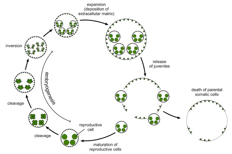
- Der asexuelle Lebenszyklus von Volvox carteri
Asexuelle männliche und weibliche Volvox-Sphäroide besitzen den gleichen Phänotyp. Unter Standardbedingungen dauert der asexuelle Lebenszyklus 48 h und wird durch einen 18 h-Hell-8 h-Dunkel-Zyklus synchronisiert. (In der Abbildung sind jeweils nur vier der sechzehn reproduktiven Zellen bzw. Embryos bzw. Tochtersphäroide eingezeichnet). Während der Embryogenese, die ca. 8 h dauert, führen die reifen reproduktiven Zellen (Gonidien) eine Serie von Zellteilungen durch (11-12 Teilungen). Eine dieser Teilungen ist asymmetrisch: Die größeren Zellen die aus dieser asymmetrischen Teilung entstehen werden zu den Gonidien der nächsten Generation, die kleineren Zellen werden später zu den somatischen Zellen. Am Ende der Teilungen befindet sich im Embryo die spätere Innenseite außen und umgekehrt, d.h. die Gonidien liegen außen und die Flagellenansätze der somatischen Zellen zeigen zum Inneren der Hohlkugel. Der gastrulationsähnliche Prozeß der Inversion, der am Ende der Embryogenese stattfindet, bringt den Embryo in die Konfiguration der erwachsenen Algen. Anschließend expandieren die Embryos, bzw. jetzt Tochtersphäroide genannt, durch den Aufbau der extrazellulären Matrix und schlüpfen aus dem elterlichen Sphäroid (="Release"). Der leere elterliche Sphäroid besteht nur aus somatischen Zellen, die nicht in der Lage sind sich weiter zu teilen. Diese somatischen Zellen altern und sterben ab. Unterdessen reifen die Gonidien der freigesetzten Tochterkolonien und beginnen wieder mit den Teilungen.
- Asexuelle und sexuelle Entwicklung in Volvox carteri

Abbildung aus: Hallmann, A., Godl, K., Wenzl, S. & Sumper, M. (1998). Trends Microbiol. 6, 185-189. Im Bild zu sehen...
-
Links: Asexueller, weiblicher Sphäroid mit 16 reproduktiven Zellen (Gonidien) und 2000-4000 somatischen Zellen. Ein asexueller, männlicher Sphäroid besitzt den gleichen Phänotyp.
-
Mitte: Sexueller, weiblicher Sphäroid mit ca. 32 Eizellen und 2000-4000 somatischen Zellen.
-
Rechts: Sexueller, männlicher Sphäroid mit ca. 128 Spermienpaketen und ca. 128 somatischen Zellen.
Volvox ist in der Lage sich sowohl asexuell als auch sexuell zu vermehren. Die sexuelle Vermehrung wird durch ein Glykoprotein-Pheromon eingeleitet, das bis zu einer Konzentration von ca. 10-16 M (!!!) volle biologische Aktivität zeigt. Somit kann ein einziges sexuelles Männchen genügend Sex-induzierendes Pheromon produzieren um viele Millionen asexueller weiblicher Sphäroide in den sexuellen Zyklus zu bringen. Das Sex-induzierende Pheromon bewirkt, daß sich das Teilungsmuster einer reproduktiven Zelle (Gonidium) ändert. Im asexuellen Zyklus von weiblichen und männlichen Volvox findet die erste asymmetrische Teilung bei 16 Zellen im 32-Zell-Embryo statt, wodurch 16 Zellen entstehen, die sich später zu Gonidien entwickeln. In Gonidien von weiblichen Sphäroiden mit Pheromon-Kontakt ist die erste asymmetrische Teilung um eine Runde verzögert, wodurch 32 Zellen entstehen, die sich später zu den 32 Eiern entwickeln. Bei Gonidien von männlichen Sphäroiden mit Pheromon-Kontakt findet die erste asymmetrische Teilung erst im 256-Zell-Embryo statt, wodurch somatische und reproduktive (sogenannte "Androgonidien") Zellen im Verhältnis 1:1 entstehen. Die Androgonidien entwickeln sich später zu Paketen aus begeißelten Spermienzellen.
-
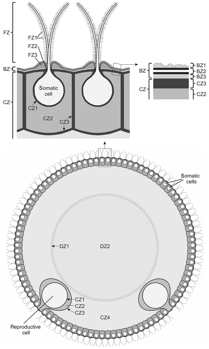
- Ein schematischer Querschnitt durch einen Volvox carteri -Sphäroid
Schematischer Querschnitt durch einen Volvox-Sphäroid unter besonderer Berücksichtigung der extrazellulären Matrix. Die extrazelluläre Matrix von Volvox ist in vier Hauptzonen unterteilt (vgl. Kirk et al., 1986; J. Cell Sci. 80, 207-231): CZ, "cellular zone", Zell-Zone; DZ, "deep zone", Innen-Zone; FZ, "boundary zone", Grenz-Zone; FZ, "flagellar zone", Flagellen-Zone.
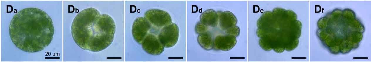
- Teilungsstadien in der Embryogenese von Volvox carteri
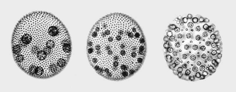
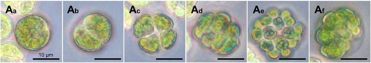
Abbildung aus: Hallmann, A. (2006). Morphogenesis in the Family Volvocaceae: Different Tactics for Turning an Embryo Right-side Out. Protist 157: 445-461. Division stages during embryogenesis of Volvox carteri. The first five cleavage stages in the development from a single reproductive cell to a 32-celled embryo are compared. D. Volvox carteri: D a, highly vacuolated gonidium prior to the onset of cleavage; D b, two-cell embryo; vacuoles have disappeared, the first cleavage furrow is visible and also the vesicle has become apparent; D c, four-cell embryo; D d, eight-cell embryo; there are two staggered rings with four cells each; the pore in the centre is the phialopore; D e, sixteen-cell embryo, D f, thirty-two-cell embryo. Scale bars are given on all pictures, and their lengths are given on the first picture.
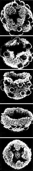
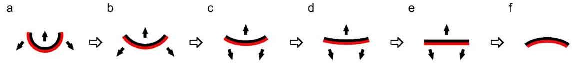
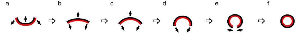
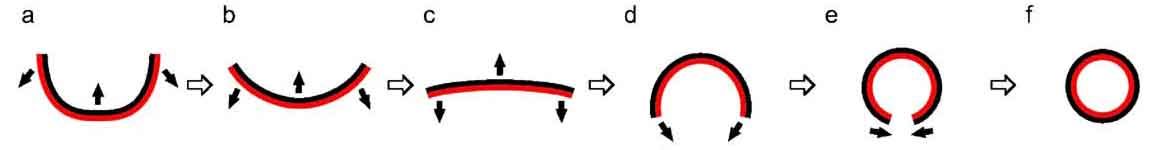
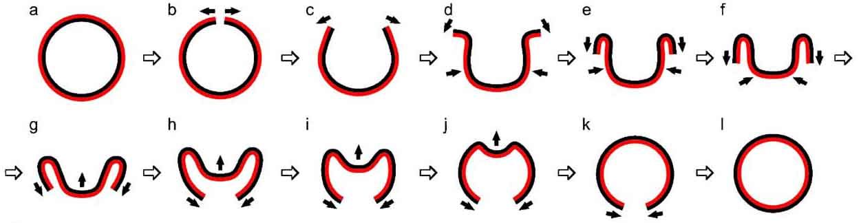
- Die Inversion eines Volvox carteri-Embryos
Während der Embryonalentwicklung von Volvox ergeben elf oder zwölf schnelle und synchrone Zellteilungen einer reproduktiven Zelle (Gonidium) sämtliche Zellen eines erwachsenen Organismus. Die entstehenden Embryonalzellen sind bereits als Hohlkugel angeordnet, aber ihre Orientierung bezüglich der Kugeloberfläche ist entgegengesetzt zu der die man in einem erwachsenen Organismus findet: Die Zellenden mit den späteren Flagellen zeigen zum Zentrum der Kugel und die reproduktiven Zellen (Gonidien) stehen von der Oberfläche ab. Während der Inversion stülpt sich der Embryo komplett um (durch einen kreuzförmigen Schlitz, die "Phialopore"), wodurch er die Orientierung der erwachsenen Organismen erhält. Die Inversion ergibt sich aus einer Folge von Zellformänderungen, die sich wellenartig von der Phialopore ausgehend zum entgegengesetzten Pol ausbreiten (siehe Abbildungen von oben nach unten).
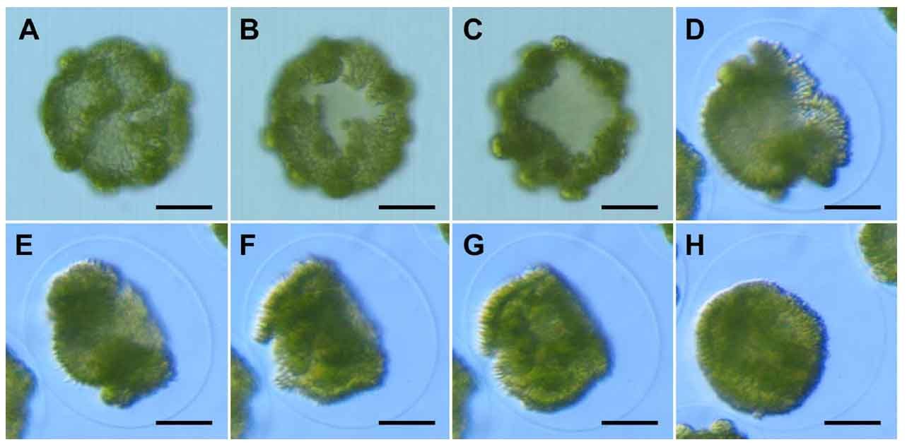
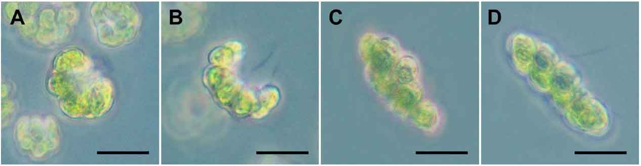
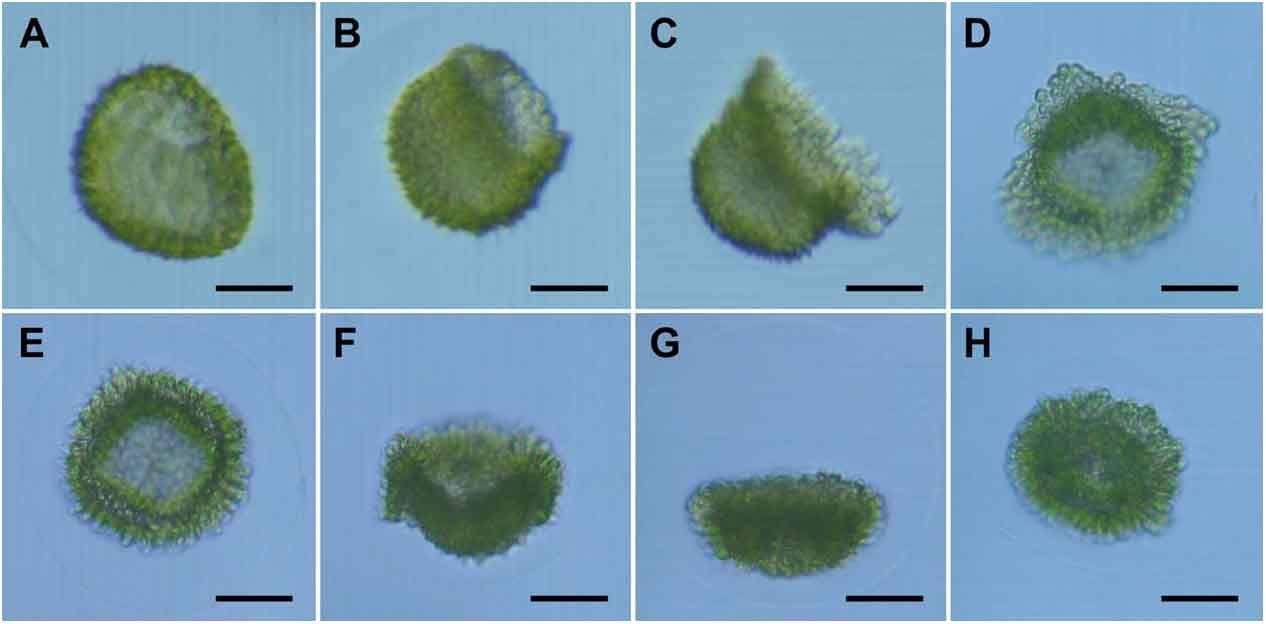
Abbildung aus: Hallmann, A. (2006). Morphogenesis in the Family Volvocaceae: Different Tactics for Turning an Embryo Right-side Out. Protist 157: 445-461. Inversion in Volvox carteri. A-H: Successive steps of inversion. Scale bars = 25 µm.
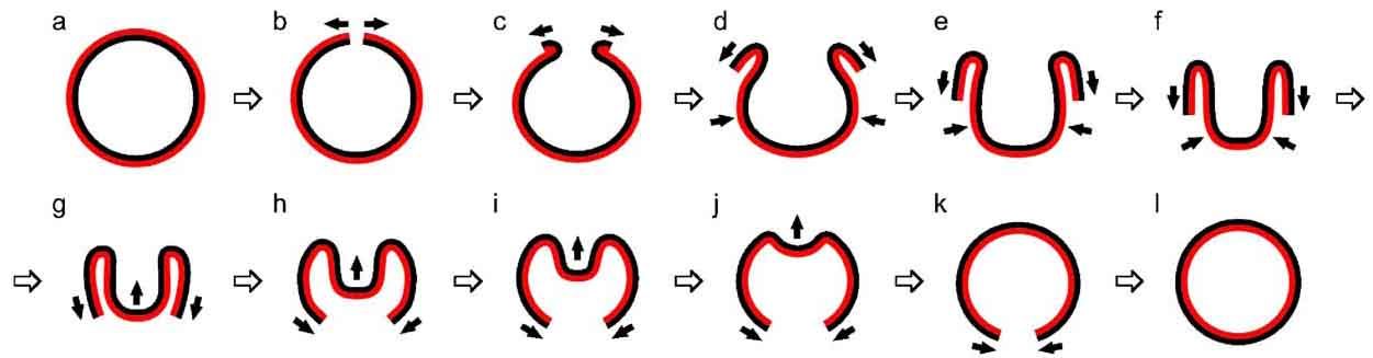
Abbildung aus: Hallmann, A. (2006). Morphogenesis in the Family Volvocaceae: Different Tactics for Turning an Embryo Right-side Out. Protist 157: 445-461. Stylized sequence of the inversion process in Volvox carteri. Cross-sections are shown. Inversion happens along the anterior-posterior axis of each organism and the anterior-posterior axis within this figure is parallel to the printed page. At the beginning of inversion, the anterior pole is at the top, after inversion the pole is on the bottom, but now (by convention) it has become the posterior pole of the inverted embryo. The side of the cell layer that is outside in the adult configuration and from which the flagella will emerge is given in black. Areas and directions of cell layer movement are indicated by small black arrows.
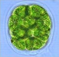
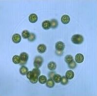
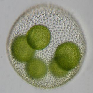
Chlamydomonas reinhardtii

Gonium pectorale
Gonium pectorale ist eine rautenförmige Kolonie mit nur einer Zellschicht aus ~16 Zellen, die durch eine gelatineartige Extrazelluläre Matrix zusammen gehalten werden.
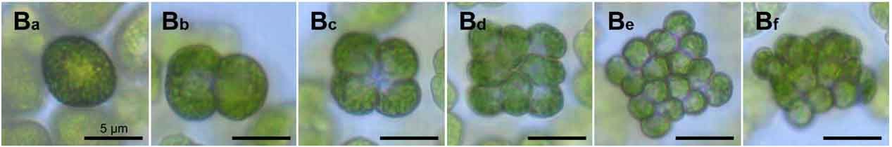
- Teilungsstadien in der Embryogenese von Gonium pectorale
Abbildung aus: Hallmann, A. (2006). Morphogenesis in the Family Volvocaceae: Different Tactics for Turning an Embryo Right-side Out. Protist 157: 445-461. Division stages during embryogenesis of Gonium pectorale. The first five cleavage stages in the development from a single reproductive cell to a 16-celled embryo are compared. A. Gonium pectorale. A a: cell prior to the onset of cleavage; A b-e: in 4 longitudinal cell cleavages a 16-cell plakea is formed; A f: side view of the 16-cell plakea showing that it is strongly curved. Scale bars are given on all pictures, and their lengths are given on the first picture.
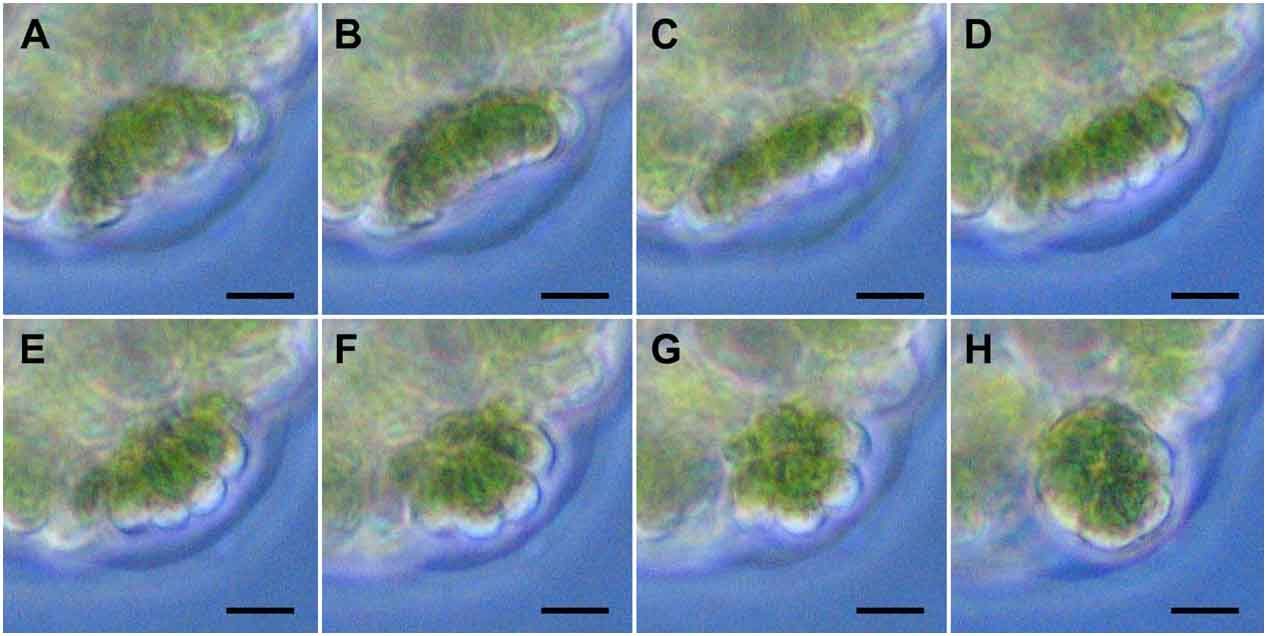
- Die Inversion eines Gonium pectorale-Embryos
Abbildung aus: Hallmann, A. (2006). Morphogenesis in the Family Volvocaceae: Different Tactics for Turning an Embryo Right-side Out. Protist 157: 445-461. Inversion in Gonium pectorale. A-D: Successive steps of inversion. Scale bars = 25 µm.
Abbildung aus: Hallmann, A. (2006). Morphogenesis in the Family Volvocaceae: Different Tactics for Turning an Embryo Right-side Out. Protist 157: 445-461. Stylized sequence of the inversion process in Gonium pectorale. Cross-sections are shown. Inversion happens along the anterior-posterior axis of each organism and the anterior-posterior axis within this figure is parallel to the printed page. At the beginning of inversion, the anterior pole is at the top, after inversion the pole is on the bottom, but now (by convention) it has become the posterior pole of the inverted embryo. The side of the cell layer that is outside in the adult configuration and from which the flagella will emerge is given in black. Areas and directions of cell layer movement are indicated by small black arrows.
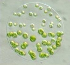
Pandorina morum
Pandorina morum ist eine ellipsoide Kolonie mit ~16 Zellen die dicht zusammen gedrängt sind und nur von relativ wenig Extrazellulärer Matrix umgeben sind.
- Teilungsstadien in der Embryogenese von Pandorina morum
Abbildung aus: Hallmann, A. (2006). Morphogenesis in the Family Volvocaceae: Different Tactics for Turning an Embryo Right-side Out. Protist 157: 445-461. Division stages during embryogenesis of Pandorina morum. The first five cleavage stages in the development from a single reproductive cell to a 32-celled embryo are compared. B. Pandorina morum. B a: cell prior to the onset of cleavage; B b-e: in 4 longitudinal cell cleavages a 16-cell plakea is formed; Bf: side view of the 16-cell plakea showing that it is curved. Scale bars are given on all pictures, and their lengths are given on the first picture.
- Die Inversion eines Pandorina morum-Embryos
Abbildung aus: Hallmann, A. (2006). Morphogenesis in the Family Volvocaceae: Different Tactics for Turning an Embryo Right-side Out. Protist 157: 445-461. Inversion in Pandorina morum. A-H: Successive steps of inversion. Scale bars = 25 µm.
Abbildung aus: Hallmann, A. (2006). Morphogenesis in the Family Volvocaceae: Different Tactics for Turning an Embryo Right-side Out. Protist 157: 445-461. Stylized sequence of the inversion process in Pandorina morum. Cross-sections are shown. Inversion happens along the anterior-posterior axis of each organism and the anterior-posterior axis within this figure is parallel to the printed page. At the beginning of inversion, the anterior pole is at the top, after inversion the pole is on the bottom, but now (by convention) it has become the posterior pole of the inverted embryo. The side of the cell layer that is outside in the adult configuration and from which the flagella will emerge is given in black. Areas and directions of cell layer movement are indicated by small black arrows.
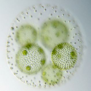
Eudorina unicocca
Eudorina unicocca ist ein kugelförmige Kolonie mit ~32 Zellen, die sich in einer relativ voluminösen Extrazellulären Matrix befinden.
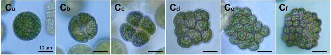
- Teilungsstadien in der Embryogenese von Eudorina unicocca
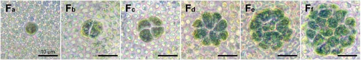
Abbildung aus: Hallmann, A. (2006). Morphogenesis in the Family Volvocaceae: Different Tactics for Turning an Embryo Right-side Out. Protist 157: 445-461. Division stages during embryogenesis of Eudorina unicocca. The first five cleavage stages in the development from a single reproductive cell to a 32-celled embryo are compared. C. Eudorina unicocca. C a: cell prior to the onset of cleavage; C b-f: in 5 cell cleavages a 32-cell curved plakea is formed. Scale bars are given on all pictures, and their lengths are given on the first pictur
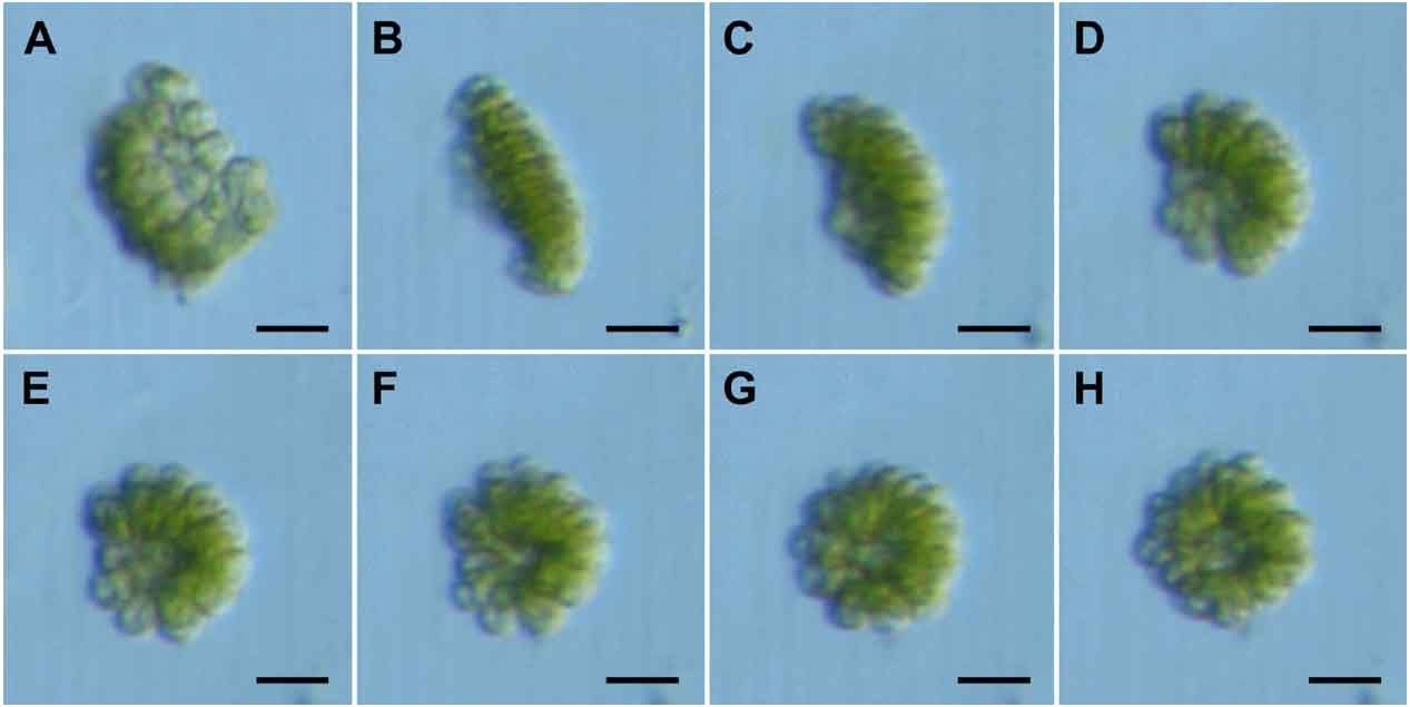
- Die Inversion eines Eudorina unicocca-Embryos
Abbildung aus: Hallmann, A. (2006). Morphogenesis in the Family Volvocaceae: Different Tactics for Turning an Embryo Right-side Out. Protist 157: 445-461. Inversion in Eudorina unicocca. A-H: Successive steps of inversion. Scale bars = 25 µm.
Abbildung aus: Hallmann, A. (2006). Morphogenesis in the Family Volvocaceae: Different Tactics for Turning an Embryo Right-side Out. Protist 157: 445-461. Stylized sequence of the inversion process in Eudorina unicocca. Cross-sections are shown. Inversion happens along the anterior-posterior axis of each organism and the anterior-posterior axis within this figure is parallel to the printed page. At the beginning of inversion, the anterior pole is at the top, after inversion the pole is on the bottom, but now (by convention) it has become the posterior pole of the inverted embryo. The side of the cell layer that is outside in the adult configuration and from which the flagella will emerge is given in black. Areas and directions of cell layer movement are indicated by small black arrows.
Pleodorina californica
Volvox tertius
Volvox tertius besteht aus ~1000 kleinen, terminal differenzierten somatischen Zellen an der Oberfläche des Sphäroiden und 4-8 Tochtersphäroiden im Inneren.
- Teilungsstadien in der Embryogenese von Volvox tertius
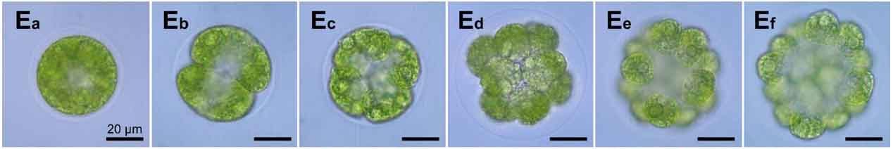
Abbildung aus: Hallmann, A. (2006). Morphogenesis in the Family Volvocaceae: Different Tactics for Turning an Embryo Right-side Out. Protist 157: 445-461. Division stages during embryogenesis of Volvox tertius. The first five cleavage stages in the development from a single reproductive cell to a 32-celled embryo are compared. E. Volvox tertius. E a, vacuolated gonidium prior to the onset of cleavage; the vesicle already detached from the gonidium; E b, two-cell embryo; E c, four-cell embryo; E d, eight-cell embryo; E e, sixteen-cell embryo, E f, thirty-two-cell embryo. Scale bars are given on all pictures, and their lengths are given on the first picture.
- Die Inversion eines Volvox tertius-Embryos
Abbildung aus: Hallmann, A. (2006). Morphogenesis in the Family Volvocaceae: Different Tactics for Turning an Embryo Right-side Out. Protist 157: 445-461. Inversion in Volvox tertius. A-H: Successive steps of inversion. Scale bars = 25 µm.
Abbildung aus: Hallmann, A. (2006). Morphogenesis in the Family Volvocaceae: Different Tactics for Turning an Embryo Right-side Out. Protist 157: 445-461. Stylized sequence of the inversion process in Volvox tertius. Cross-sections are shown. Inversion happens along the anterior-posterior axis of each organism and the anterior-posterior axis within this figure is parallel to the printed page. At the beginning of inversion, the anterior pole is at the top, after inversion the pole is on the bottom, but now (by convention) it has become the posterior pole of the inverted embryo. The side of the cell layer that is outside in the adult configuration and from which the flagella will emerge is given in black. Areas and directions of cell layer movement are indicated by small black arrows.
Volvox globator
Volvox globator besteht aus ~3000 kleinen, terminal differenzierten somatischen Zellen an der Oberfläche des Sphäroiden und 4-6 Tochtersphäroiden im Inneren.
- Teilungsstadien in der Embryogenese von Volvox globator
Abbildung aus: Hallmann, A. (2006). Morphogenesis in the Family Volvocaceae: Different Tactics for Turning an Embryo Right-side Out. Protist 157: 445-461. Division stages during embryogenesis of Volvox globator. The first five cleavage stages in the development from a single reproductive cell to a 32-celled embryo are compared. F. Volvox globator. F a, vacuolated gonidium prior to the onset of cleavage (surrounded by somatic cells); F b, two-cell embryo; F c, four-cell embryo; F d, eight-cell embryo; F e, sixteen-cell embryo, F f, thirty-two-cell embryo. Scale bars are given on all pictures, and their lengths are given on the first picture.
- Die Inversion eines Volvox globator-Embryos
Abbildung aus: Hallmann, A. (2006). Morphogenesis in the Family Volvocaceae: Different Tactics for Turning an Embryo Right-side Out. Protist 157: 445-461. Inversion in Volvox globator. A-H: Successive steps of inversion. Scale bars = 25 µm.
Abbildung aus: Hallmann, A. (2006). Morphogenesis in the Family Volvocaceae: Different Tactics for Turning an Embryo Right-side Out. Protist 157: 445-461. Stylized sequence of the inversion process in Volvox globator. Cross-sections are shown. Inversion happens along the anterior-posterior axis of each organism and the anterior-posterior axis within this figure is parallel to the printed page. At the beginning of inversion, the anterior pole is at the top, after inversion the pole is on the bottom, but now (by convention) it has become the posterior pole of the inverted embryo. The side of the cell layer that is outside in the adult configuration and from which the flagella will emerge is given in black. Areas and directions of cell layer movement are indicated by small black arrows.