Bielefeld Institute for Biophysics and Nanoscience - Methodes of Analysis

Introduction
Preparation
- Ultra-high vacuum electron beam evaporation
- Metal organic vapour phase deposition (MOCVD)
- Nanometer and micrometer lateral structuring
- Chemical vapour deposition (CVD)
- Physical vapour deposition (PVD)
- Lithography in nanometer and micrometer structures, laser and electron beam writing and reactive ion etching (RIE)
- Organic and inorganic (metallic and oxide) mono- and multilayers in the nm range
- Langmuir film balance
Microscopy
- Light microscopy (dark and bright field, phase contrast)
- Confocal laser scanning microscope (LSM)
- High-resolution fluorescence microscopy (FM, dStorm)
- 3D structured illumination microscopy (3D-SIM)
- Transmission electron microscopy (TEM)
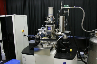
- Scanning electron microscopy (SEM) with EDX
- Photoemission electron microscopy (PEEM)
- Helium ion microscopy (HIM)
- Scanning probe microscopy and force spectroscopy (AFM, STM, MFM) in air, in liquids and in ultra-high vacuum (UHV)
- Scanning near-field optical microscopy (SNOM)
Spectroscopy
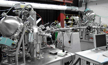
- Secondary ion mass spectroscopy (SIMS)
- UV photoelectron spectroscopy (UPS)
- X-ray photoelectron spectroscopy (XPS)
- Raman spectroscopy
- Transient absorption spectroscopy with ns time resolution and optical coincidence analyses
- Time-resolved fluorescence spectroscopy with picosecond time resolution
Further methods of analysis
- X-ray diffraction (small-angle and large-angle diffraction)
- X-ray reflectivity
- Ellipsometry
- Contact angle measurements
- Alternating gradient magnetometer
- Electrical and magneto-transport measurements, magnetometry
- Magneto-optical Kerr effect
- High-pressure liquid chromatography (HPLC)
- Electrophoresis
- Optical tweezers (OT) in single and multiple beams
- Capillary electrophoresis in microchips (Integrated CE)

